NCERT Solved Exercise Questions – Class 11 Biology Chapter 7 Structural Organisation in Animals
7.1 Answer in one word or one line.
(i) Give the common name of Periplanata americana.
(ii) How many spermathecae are found in earthworm?
(iii) What is the position of ovaries in cockroach?
(iv) How many segments are present in the abdomen of cockroach?
(v) Where do you find Malpighian tubules?
Ans – (i) Cockroach
(ii) Four pairs of spermathecae are found in the 6“ to 9th segments (one pair in each segment).
(iii) The abdomen of the cockroach has two ovaries that are located laterally in the second to sixth abdominal segments.
(iv) ten
(v)At the intersection of the midgut and hindgut,. Malpighian tubules are 100–150 yellow, thin filamentous rings that are found in earthworms.
7.2 Answer the following:
(i) What is the function of nephridia?
(ii) How many types of nephridia are found in earthworm based on their location?
Ans – (i) Earthworms have nephridia, which serve as their excretory and osmoregulating organs. The bodily fluids’ volume and make-up are controlled by nephrodia. A nephridium is a small, coil-like structure that initially functions as a funnel to collect extra fluid from the coelomic chamber. Through a pore in the body wall or into the digestive tube, the funnel is connected to a tubular waste stream.
(ii) Nephridia are found in all segments of the earthworm, with the exception of the first two. Nephridia can be divided into three categories based on where they are found:
(a)From segment 15 until the final segment that opens into the colon, there are septal nephridia on both sides of the intersegmental septa.
(b) Integumentary nephridia that are attached to the body wall’s lining from segment three to the last segment and open on the body surface.
(c) In the fourth, fifth, and sixth segments, there are three paired tufts of pharyngeal nephridia.
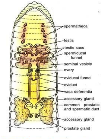
7.3 Draw a labelled diagram of the reproductive organs of an earthworm.
Ans –

7.4 Draw a labelled diagram of alimentary canal of a cockroach.
Ans –

7.5 Distinguish between the followings
(a) Prostomium and peristomium
(b) Septal nephridium and pharyngeal nephridium
Ans –
(a) Differences between prostomium and peristomium are
| Prostomium | Peristomium |
| A small, fleshy lobe called a protomium hangs over an earthworm’s mouth. It is sensory in nature and aids the organism in pushing into the soil. | The peristomium is the first part of the earthworm’s body. It encircles the gaping mouth. |
(b) Differences between septal and pharyngeal nephridia are:
| Septal nephridium | Pharyngeal nephridium |
| They open into the intestine and are located on both sides of the inter-segmental septa behind the 15th segment. | They are present as three paired tufts in the fourth, fifth, and sixth segments. |
7.6 What are the cellular components of blood?
Ans – Erythrocytes (RBCs), leucocytes (WBCs), and thrombocytes are blood constituents (platelets). These elements make up 45% of blood. In the plasma, the remaining fluid, they are suspended. Erythrocytes from mammals are biconcave, coloured cells without nuclei. They support the movement of respiratory gases. White blood cells, or leucocytes, have nuclei. Granulocytes (neutrophils, eosinophils, and basophils) and agranulocytes are the two categories into which they can be separated (lymphocytes and monocytes). They support the body’s defences against numerous disease-causing substances. Cell fragments called thrombocytes are created from bone megarkaryocytes. They are very important for blood coagulation.
7.7 What are the following and where do you find them in animal body.
(a) Chondriocytes
(b) Axons
(c) Ciliated epithelium
Ans – (a) Chondrocytes are the only cells that may be detected in cartilage . They create and preserve the cartilage matrix while being present in regions known as lacunae. Chondrocytes are responsible for cartilage’s ability to bend. The epiglottis, pinna, and tip of the nose all have cartilage.
(b) Axon – An axon is one of a neuron’s processes, which is the structural and operational component of the nervous system. Axons begin in the region of the cell called the cyton from which they emerge, and they end in a clump of branches known as terminal arborizations. It carries electrical impulses out of the cyton. The brain and spinal cord both include neurons (nerve cells).
(c) Ciliated epithelium: This type of epithelium is made up of columnar or cuboidal cells that have cilia on their free surfaces. Their job is to move particles or mucus in a certain direction over the epithelium; they are present in the lining of the stomach and intestine and aid in secretion and absorption. The inner surfaces of hollow organs like bronchioles and fallopian tubes are where they are primarily found.
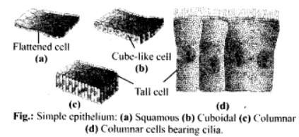
7.8 Describe various types of epithelial tissues with the help of labelled diagrams.
Ans – The inner and exterior lining of numerous organs are covered by epithelial tissues. There is little intercellular matrix between the tightly packed cells of epithelial tissues.
Epithelial tissues come in two varieties:
(i) Simple epithelium
(ii) Compound epithelium
(i) Simple epithelium :
consists of a single layer of cells and serves as the lining for ducts, tubes, and bodily cavities. By modifying the structure, it is further separated into three types:
(A) Squamous epithelium: It is made up of a single layer of flat cells that are closely packed together and have an oval or spherical nucleus in the centre. Another name for it is pavement epithelium. It is present in the lining of eye lenses, the walls of blood vessels, and the air sacs of the lungs.
(b) Cuboidal epithelium: The nucleus of cuboidal epithelium cells is centred and each cell is as tall as it is wide. Secretion and absorption are its primary activities. It lines the salivary, thyroid, and sweat glands. The proximal portion of the uriniferous tubule, pancreatic duct, testis, and ovary are lined by brush edged cuboidal epithelium, which are cells with microvilli on their free surface.
(c) Columnar epithelium: Cells have nuclei that are basally positioned. Both secretion and absorption are enhanced. It happens in the stomach, gall bladder, and gut lining.

(d) Ciliated epithelium: Columnar and cuboidal cells have cilia covering their free surfaces. Cilia aid in directing the movement of substances such as fluids, particles, mucus, etc. It develops on the inner surface of the bronchioles, nasal canal, and Fallopian tubules.

- Pseudostratified epithelium: This type of epithelium is composed of a single layer of cells, some of which are shorter than others. The epithelium seems to be 2-3 layers as a result of the variation in cell size. Both the parotid salivary gland and the urethra have pseudostratified columnar epithelium. In the lining layer of the nasal chambers, trachea, and large bronchi, pseudostretified columnar ciliated epithelium (only larger cells ciliated) occurs. Moving mucus and foreign objects is made easier by it.
Based on the presence or absence of duct glands, it is possible to:
(a) Exocrine glands : These glands pour their secretion through a duct. For example, goblet cells, salivary glands, tear glands, stomach glands, and intestinal glands all release milk, saliva, mucus, and earwax.
(b) Endocrine glands: These ductless glands secrete into the blood or lymph in order to reach the target area. They produce what is known as hormones, such as those produced by the pituitary, thyroid, parathyroid, and adrenal glands.
(c) Heterocrine glands, such as the pancreas, are both exocrine and endocrine.
Depending on how they secrete, glands can be:
Merocrine: Release of secretion
via diffusion, such as sweat glands and goblet cells.
(b) Apocrine glands: In the terminal portion of the cell that is severed, such as in mammary glands, glandular secretion builds up.
(c) Holocrine glands: The cell filled with secretory product, such as the sebaceous gland, disintegrates during discharge of the product.
7.9 Distinguish between
(a) Simple epithelium and compound epithelium
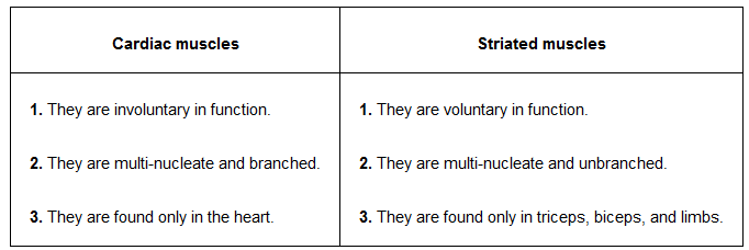
(b) Cardiac muscle and striated muscle
(c) Dense regular and dense irregular connective tissues
(d) Adipose and blood tissue
(e) Simple gland and compound gland
Ans –
- Simple epithelium and compound epithelium

(b) Differences between cardiac and striated muscles are as follows:

The following are the main distinctions between dense regular connective tissues and dense irregular connective tissues:.

The main difference between adipose tissue and blood tissue are as following.

The main difference between simple gland and compound gland are as following

7.10 Mark the odd one in each series:
(a) Areolar tissue; blood; neuron; tendon
(b) RBC; WBC; platelets; cartilage
(c) Exocrine; endocrine; salivary gland; ligament
(d) Maxilla; mandible; labrum; antennae
(e) Protonema; mesothorax; metathorax; coxa
Ans – (a) Neuron: Blood, tendon, and areolar tissue are connective tissues, whereas neurons are a component of nervous tissues.
(b) Cartilage: Whereas platelets, RBCs, and WBCs are a part of vascular connective tissue, cartilage is a part of the skeleton.
(c) Ligament: Ligament is a type of connective tissue.
(d) Antennae: The cockroach’s antennae are sensory organs, but its maxilla, mandible, and labrum are mouth parts.
(e) Protonema: A juvenile filamentous stage in the life cycle of Bryophytes, whereas the mesothorax, metathorax, and coxa are cockroach appendages.
7.11 Match the terms in column I with those in column II:
Column I Column II
(a) Compound epithelium (i) Alimentary canal
(b) Compound eye (ii) Cockroach
(c) Septal nephridia (iii) Skin
(d) Open circulatory system (iv) Mosaic vision
(e) Typhlosole (v) Earthworm
(f) Osteocytes (vi) Phallomere
(g) Genitalia (vii) Bone
Ans – (a) – (iii)
(b) – (iv)
(c) – (v)
(d) – (ii)
(e) – (i)
(f) – (vii)
(g) – (vi)
7.12 Mention breifly about the circulatory system of earthworm
Ans – As the blood flows through closed blood vessels, earthworms have a closed form of blood vascular system. Because haemoglobin is a respiratory pigment, blood is red in colour. Dorsal, ventral, sub-neural, lateral oesophageal, and supra-oesophageal blood arteries are prominent in earthworms. There are four valve-equipped tubular heart pairs. The dorsal and ventral blood vessels are connected by the anterior two pairs of hearts, or lateral hearts, which are located in the seventh and ninth segments, respectively. They transport blood from the ventral blood vessel to the dorsal blood vessel. The 12th and 13th segments include the posterior two pairs of hearts known as latero-oesophageal hearts.
In addition to joining the dorsal and ventral blood vessels, the latero-oesophageal hearts also link with the supra oesophageal blood vessel.
Blood is transported to the ventral blood vessel by lateral oesophageal hearts from the dorsal vessel and the supra oesophageal vessel. Blood continues to flow in one direction due to contractions. The 4th, 5th, and 6th segments of the HLH contain blood glands that create blood cells and haemoglobin, which is dissolved in blood plasma. The nature of blood cells is phagocytic.
7.13 Draw a neat diagram of digestive system of frog.
Ans –

7.14 Mention the function of the following
(a) Ureters in frog
(b) Malpighian tubules
(c) Body wall in earthworm
Ans – (a) Ureters in frog: Ureter is a transparent duct which arise from outer portion of kidney. The ureter serves as the urinogenital duct in male frogs, running backward from the kidneys and opening into the cloaca. It transports spermatozoa and urine from the kidney to the cloaca. Only urine is transported by the female ureter from the kidneys to the cloaca.
(b) Malpighian tubules:
1. They are a cockroach’s excretory system, to start with.
2. They take the nitrogenous excretions out of the haeomolymph and deliver them to the intestine.
3. Cells with glands and cilia border each tubule. They take up nitrogenous wastes and transform them into uric acid, which is then eliminated through the hindgut.
(c) The cuticle, epidermis, muscle layer, and parietal peritoneum make up the earthworm body wall.
(i) It preserves the body’s distinctive shape.
(ii) It safeguards the interior organs .
(iii) The cuticle limits excessive evaporation .
(iv) It is the perfect respiratory organ.
(v) The receptor cells perform an essential sensory role.
(vi) The albumen aids in the cocoon’s development. The developing earthworm inside the cocoon uses it as nourishment as well.
(vii) The muscles and setae are in charge of propulsion.
(viii). Nephridiopores allow excretory materials to leave the body


