NCERT Solved Exercise Questions – Class 11 Biology Chapter 6 Anatomy of Flowering Plants
6.1 State the location and function of different types of meristems.
Ans – Based on where they reside in the plant body, meristems can be divided into three categories:
(i) Apical meristem: This structure can be found at the tips of roots and shoots. At the tip of the shoots is the shoot apical meristem, which divides actively to lengthen the stem and produce new leaves. Root elongation is assisted by the root apical meristem.
(ii) The intercalary meristem,: which can be found at the bases of leaves above or below the nodes and is in charge of the organs’ elongation, is a part of the leaf.
(iii) Lateral meristem: Located on the lateral side, it is in charge of increasing diameter or girth.
6.2 Cork cambium forms tissues that form the cork. Do you agree with this statement? Explain.
Ans – Another meristematic tissue known as cork cambium or phellogen eventually forms, typically in the cortical region. The cork cambium is made up of several layers. It is constructed of compact, almost rectangular compartments with thin walls. Cells on both sides are cut off by the cork cambium. While the inner cells evolve into secondary cortex or phelloderm, the outer cells become cork or phellem. Water cannot penetrate the cork because to suberin deposition in the cell wall. The secondary cortex’s cells are parenchymatous. Periderm is the aggregate name for phellogen, phellem, and phelloderm.
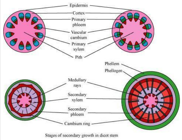
6.3 Explain the process of secondary growth in the stems of woody angiosperms with the help of schematic diagrams. What is its significance?
Ans – The interfascicular cambium is the strip of cambium that exists between the primary xylem and phloem in woody dicots. The cells of the medullary rays that are next to the interfascicular cambium create the interfascicular cambium. As a result, a continuous cambium ring develops. New cells are cut off from the cambium’s sides. While the cells cut off toward the pith give rise to the secondary xylem, the cells present toward the outside differentiate into the secondary phloem. There is a greater production of secondary xylem than secondary phloem.

1. The action of the vascular cambium and cork cambium in stellar and extrasolar regions is the cause of its “permanent expansion in thickness.” There is intra fascicular cambium in dicot stems.
2. The medullary ray cells undergo meristematic change and develop into interfascicular cambium.
3. By joining together, these two cambiums form a complete cambial ring.
4. On both its inner and exterior sides, its cells divide to create new cells.
5. The inner side’s cells differentiate into secondary xylem, whilst the outer side’s cells divide into secondary phloem.
6. As the plant’s stem becomes larger, the epidermis is replaced with a secondary protective tissue. The material is phellogen (cork cambium).
6.4 Draw illustrations to bring out the anatomical difference between
(a) Monocot root and Dicot root
(b) Monocot stem and Dicot stem
Ans –


6.5 Cut a transverse section of young stem of a plant from your school garden and observe it under the microscope. How would you ascertain whether it is a monocot stem or a dicot stem? Give reasons.
Ans – In monocot stems, vascular bundles are dispersed throughout the ground tissue, whereas in dicot stems, vascular bundles are organised in a ring. It is possible to determine whether a young stem is a dicot or a monocot based on the configuration of the vascular bundles. Other distinguishing characteristics of monocot stems include sclerenchymatous hypodermis, oval or circular vascular bundles, and Y-shaped xylem in addition to undifferentiated ground tissue.
6.6 The transverse section of a plant material shows the following anatomical features –
(a) the vascular bundles are conjoint, scattered and surrounded by a sclerenchymatous bundle sheaths.
(b) phloem parenchyma is absent. What will you identify it as?
Ans – A typical juvenile monocotyledonous stem’s transverse section reveals that
- Sclerenchymatous bundle sheaths cover the conjoint, dispersed, and vascular bundles.
2. The vascular bundles lack phloem parenchyma and have water-containing holes.
6.7 Why are xylem and phloem called complex tissues?
Ans – Given that they are composed of several cell types, xylem and phloem are referred to as complex tissues. To carry out the numerous xylem and phloem duties, these cells cooperate and act as a unit.
Xylem aids in the transportation of minerals and water. Additionally, it gives plants mechanical assistance. It consists of the following elements:
• Tracheids (xylem vessels and xylem tracheids)
• Xylem parenchyma
• Xylem fibres
Dead cells with thick walls and tapered ends are called tracheids. The vessel members combine to form long, tubular, cylindrical structures called vessels. Each vessel has lignified walls and a sizable centre cavity. Protoplasm is absent from arteries and tracheids. Xylem fibres have substantial wall thickness and a little lumen. They assist in giving the plant mechanical support. Thin-walled parenchymatous cells, which make up the xylem parenchyma, aid in the radial conduction of water and the storage of food resources.
Phloem aids in transporting meal components. It is made up of:
• Sieve tube elements
• Companion cells
• Phloem parenchyma
• Phloem fibres
6.8 What is stomatal apparatus? Explain the structure of stomata with a labelled diagram.
Ans – The epidermis of leaves contains stomata, or pores. The exchange of gases and the process of transpiration are regulated by stomata. Two bean-shaped cells called guard cells, which enclose the stomatal pore, make up each stoma. Guard cells have thin outer walls that face away from the stomatal pore and thick inner walls that face the pore. The guard cells control stomata opening and closing and have chloroplasts. Sometimes, a small number of epidermal cells nearby the guard cells specialise in their size and shape and are referred to as subsidiary cells.
- All of a plant’s green aerial parts have a few tiny openings called stomata on their epidermis, but they are particularly numerous on the bottom surface of the leaves because they control the transpiration process.
2.The upper surface of aquatic plant leaves have a significant number of stomata.
3.The guard cells, which surround each stomata, are two separate cells. These are bean-shaped in dicotyledon plants, but dumb-bell-shaped in sedges and grasses.
4. There is life in the guard cell. Their inner walls, which encircle the hole, are far thicker than their thin exterior walls.
6.9 Name the three basic tissue systems in the flowering plants. Give the tissue names under each system.
Ans – There are three different types of tissue systems based on their location and structural characteristics. This is the
the vascular or conducting tissue system, the ground or fundamental tissue system, and the epidermal tissue system.
1. The epidermal tissue network The epidermal tissue system, which is made up of epidermal cells, stomata, and the epidermal appendages trichomes and hairs, forms the outermost layer of the entire plant body.
2. The ground tissue is made up of all tissues, excluding the epidermis and vascular bundles. Simple tissues like parenchyma, collenchyma, and sclerenchyma make up this tissue type.
3.Phloem and xylem, two complex tissues, make up the vascular system. Vascular bundles are composed of both the xylem and the phloem.
6.10 How is the study of plant anatomy useful to us?
Ans – Understanding the structural adaptations of plants in relation to various environmental situations is made possible by the study of plant anatomy. Additionally, it aids in the differentiation of gymnosperms, dicots, and monocots. The physiology of plants is related to such a study. As a result, it aids in improving food crops. We can forecast the strength of wood thanks to the research of plant structure. This is helpful in making the most of it. The commercial exploitation of different plant fibres like jute, flax, and others is aided by research into those fibers.
6.11 What is periderm? How does periderm formation take place in the dicot stems?
Ans – Periderm is the aggregate name for phellogen, phellem, and phelloderm. In the cortical region, phellogen typically forms. There are a few levels of phellogen. It is constructed of compact, almost rectangular compartments with thin walls. Cells are severed on both sides by phellogen. While the inner cells evolve into secondary cortex or phelloderm, the outer cells become cork or phellem. Water cannot penetrate the cork because to suberin deposition in the cell wall. The secondary cortex’s cells are parenchymatous.
6.12 Describe the internal structure of a dorsiventral leaf with the help of labelled diagrams
Ans – Dicots are known to have dorsiventral leaves. There are three separate portions to a dorsiventral leaf’s vertical segment.
(1) The epidermis
Both the top surface (adaxial epidermis) and the bottom surface have epidermis (abaxial epidermis). A thick cuticle surrounds the epidermis on the exterior. More stomata are present in the abaxial epidermis than the adaxial epidermis.
[2] Mesophyll:
A tissue of the leaf called mesophyll is found between the adaxial and abaxial epidermises. It is divided into the spongy parenchyma and the palisade parenchyma, which are both made up of tall, tightly packed cells (comprising oval or round, loosely-arranged cells with inter cellular spaces). The chloroplasts found in mesophyll are responsible for photosynthesis.
Elongated cells that are positioned adaxially and parallel to one another make up the palisade parenchyma.
The spongy parenchyma, which is oval or spherical and loosely organised, lies below the palisade cells and reaches to the lower epidermis.
[3] Vascular system:
Vascular bundles, which are visible in the veins and the midrib, are included in this.
The veins’ diameter determines the size of the vascular bundles.
In the dicot leaves’ reticulate venation, the veins’ thickness varies. A layer of cells called bundle sheaths with thick walls surrounds the vascular bundles.


